
Figure 4.
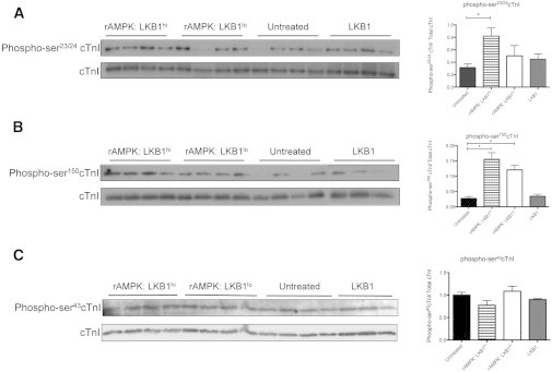
Western blot analysis of phosphorylated cTnI. The Western blot depicts phospho-cTnI in myofibrils treated with various stoichiometric LKB1 activations of AMPK and the LKB1 complex, with the corresponding normalized optical density plotted on the right. (A) Phospho-ser23/24 cTnI was measured in treated and untreated myofibrils. Fibers treated with a high level of LKB1 activation of AMPK (rAMPK/LKB1hi) had a significant increase in phosphorylated ser23/24 content relative to untreated myofibrils (p < 0.05; n = 4 for all groups). (B) Phospho-ser150 cTnI was measured in treated and untreated myofibrils. There was a significant increase in phosphorylated ser150 in rAMPK/LKB1hi- and rAMPK/LKB1lo-treated fibers (p < 0.05; n = 3–4 per group). (C) Phospho-ser43cTnI was measured in treated and untreated myofibrils. There was no change in phospho-ser43cTnI in any treatment group (p > 0.05).