
Figure 6.
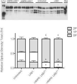
Phosphate-affinity SDS-PAGE (Phos-tag) of the total cTnI phosphospecies distribution. cTnI was separated into unphosphorylated [0P], monophosphorylated [1P], and bisphosphorylated [2P] phosphospecies by SDS-PAGE-Phos-tag followed by Western blot analysis with a total cTnI antibody. Top panel: representative SDS-PAGE-Phos-tag followed by Western blot, illustrating three bands corresponding to 0P, 1P, and 2P. Human recombinant cTnI (hcTnI) and PKA-treated (+PKA) cardiomyocytes were used to confirm each phosphospecies. Bottom panel: stacked bar graph indicating the relative amounts of each phosphospecies per experimental group. There is a significant shift in fibers treated with both high and low LKB1 activation of AMPK (rAMPK/LKB1hi and rAMPK:LKB1lo) away from the 0P state (p < 0.05). There is no change in total cTnI phosphospecies distribution in the LKB1-complex-treated group.