
Figure 2.
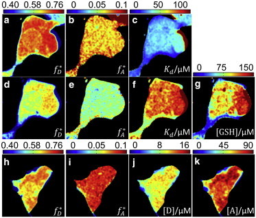
Validation of MC-FLIM-FRET in cells. (a–c) Bound fractions and dissociation constants. (d–g) Recovered parameters upon adding competitor GSH. (h–k) Absolute concentration determination in cells with known Kd = 37.2 ± 0.2 μM. The average photon count in each binned pixel is ∼13,000 (see Section S3 in the Supporting Material for details).