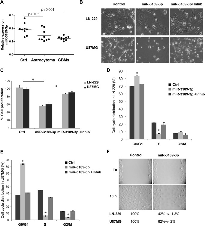
FIGURE 1.
MiR-3189-3p is down-regulated in glial tumors and affects growth and migration of glioblastoma cells in culture. A, relative expression of miR-3189-3p in controls (Ctrl), astrocytomas, and glioblastomas (GBM) (n = 9/group). B, phase-contrast images showing the morphology of LN-229 and U87MG cells following transfection with miR-3189-3p or miR-3189-3p + anti-miR-3189-3p (original magnification, ×10). Images were acquired 48 h post-transfection. Inhib, inhibitor. C, cell proliferation assay performed 72 h post-transfection of the indicated cell lines with mock, miR-3189-3p, or miR-3189-3p + inhibitor and quantified using 3-(4,5-dimethylthiazol-2-yl)-5-(3-carboxymethoxyphenyl)-2-(4-sulfophenyl)-2H-tetrazolium reagent. Results are expressed as percent of growth/mock-treated control. D and E, cell cycle analysis of LN-229 (D) and U87MG (E) cells transfected with mock (ctrl), miR-3189-3p, and anti-miR-3189-3p (Inhib). Cells were stained with Guava cell cycle reagent and cell cycle distribution (%) was quantified by flow cytometry using a FACSAria. F, representative images of a scratch assay to monitor migration of controls (mock-transfected) and miR-3189-3p-transfected LN-229 cells (original magnification, ×10). Migration into the cell-free area was monitored by time-lapse imaging in a VivaView fluorescent microscope. The same experiment was performed in U87MG cells, and data are shown below the images in F. *, p < 0.05.

