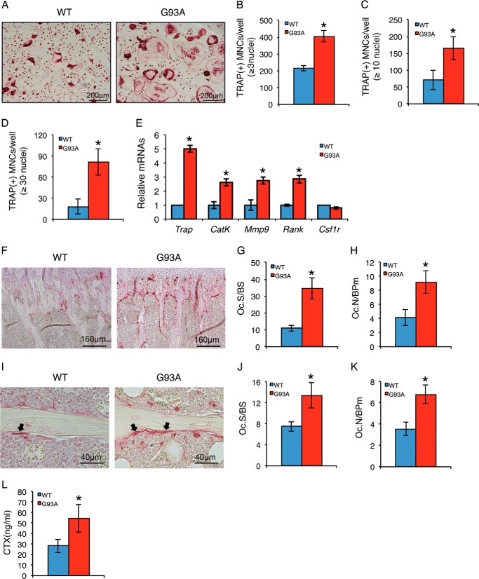
FIGURE 4.
Osteoclast formation is enhanced in primary BMM cultures and in bones of 4-month-old G93A mice with muscle atrophy. A–D, in vitro osteoclast formation. Primary BMMs isolated from 4-month-old wild type (WT) and G93A female mice were differentiated with macrophage colony stimulating factor (M-CSF) (10 ng/ml) and RANKL (50 ng/ml) for 5 days followed by TRAP staining. TRAP-positive MNCs (B, ≥3 nuclei; C, ≥10 nuclei; D, ≥30 nuclei) per well were scored (B–D). *, p < 0.05 (versus WT). Experiments were repeated three times with six samples per group. E, RT-qPCR analysis. Primary BMMs were differentiated for 5 days followed by RT-qPCR analysis using primers for Trap, Cat K, Mmp9, Rank, and Csf1r. Gapdh mRNA was used as internal control. *, p < 0.05 (versus WT). Experiments were repeated three times in quadruplicate. F–K, in vivo osteoclast formation. Tibial sections of 4-month-old WT and G93A female mice were stained for TRAP activity. Arrowheads indicate osteoclasts on trabecular surfaces. Quantitative data of Oc.S/BS (G and J) and Oc.N/BPm (H and K) in primary (F–H) and secondary (I–K) spongiosa. *, p < 0.05 (versus WT), n = 7. L, serum C-terminal telopeptide of type 1 collagen (CTX) assay. Serum CTX levels were assayed. *, p < 0.05 (versus WT), n = 7. CatK, cathepsin K; Mmp9, matrix metallopeptidase 9; Rank, receptor activator of nuclear factor κB; Csf1r, colony stimulating factor 1 receptor.

