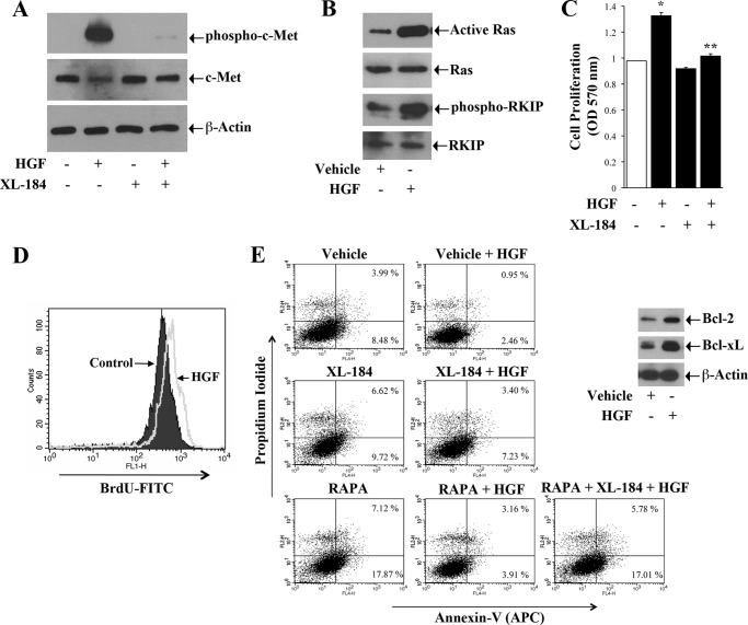
FIGURE 1.
Induction of c-Met activates Ras, promotes proliferation and inhibits apoptosis of renal cancer cells. A, 786-O cells were incubated with either XL-184 (10 μm) or vehicle alone for 2 h and then treated with either HGF (50 ng/ml) or vehicle for 15 min. Following treatment, cells were lysed, and the cell lysates were used to measure the levels of phospho-c-Met, c-Met, and β-actin (internal control) by Western blot analysis. B, 786-O cells were treated with HGF as described in A. Cell lysates were prepared utilizing a Ras activation kit as described under “Experimental Procedures,” and the expression of GTP-bound Ras was subsequently analyzed by Western blot. Cell lysates were also used to measure the expression of total Ras, phospho-RKIP, and total RKIP by Western blot. C, 786-O cells were incubated with either XL-184 (10 μm) or vehicle alone for 2 h, and then treated with either HGF (50 ng/ml) or vehicle. After 48 h of treatment, cell proliferation was measured by MTT assay. D, 786–0 cells were labeled with 10 μm BrdU, and then treated with either HGF (50 ng/ml) or vehicle alone for 24 h. Following treatment, the cells were stained with BrdU-FITC antibody and analyzed by flow cytometry. E, 786-O cells were incubated with either XL-184 (10 μm) or vehicle alone for 2 h and then treated with different combinations of HGF (50 ng/ml) and RAPA (10 ng/ml). Following 48 h of treatment, apoptotic index of the cells was determined by annexin V (APC) and propidium iodide staining. In right panel, the lysates from vehicle- and HGF-treated cells were used to measure the expression of Bcl-2, Bcl-xL, and β-actin (internal control) by Western blot. A, B, D, and E are representative of three independent experiments. C, the columns represent the mean ± S.D. of triplicate readings of two different samples. *, p < 0.05 compared with vehicle-treated control, and **, p < 0.05 compared with only HGF-treated cells.

