
FIGURE 4.
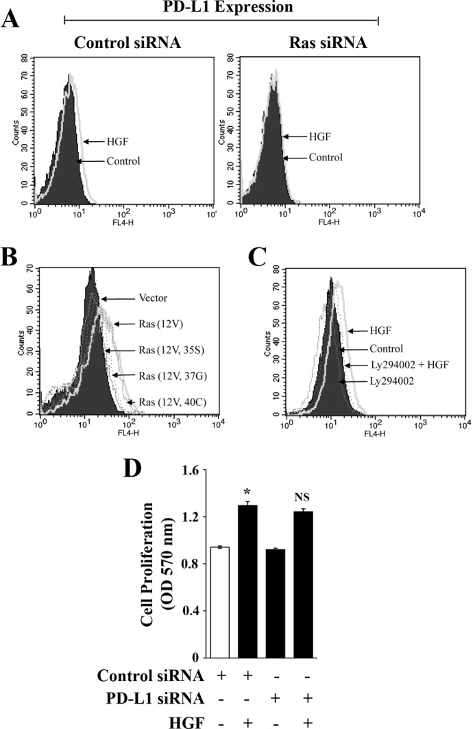
c-Met-mediated PD-L1 up-regulation is channeled through Ras-PI-3K pathway. A, 786-O cells were transfected with 50 nm of either control siRNA or Ras-siRNA. Following 48 h of transfection, cells were treated with HGF (50 ng/ml)/vehicle for an additional 36 h, and PD-L1 expression was analyzed by flow cytometry. B, 786-O cells were transfected with either one of the three Ras effector domain mutant plasmids Ras(12V, 35S), Ras(12V, 37G), Ras(12V, 40C), or active Ras plasmid Ras(12V) or empty vector alone. Following 48 h of transfection, cells were analyzed for PD-L1 expression by flow cytometry. C, 786-O cells were incubated with either LY294002 (10 μm) or vehicle for 2 h and then treated with HGF (50 ng/ml)/vehicle for 36 h. Following treatment, cells were analyzed for PD-L1 expression by flow cytometry. D, 786–0 cells were transfected with 50 nm of either control siRNA or PD-L1 siRNA. Following 48 h of transfection, the cells were treated with either HGF (50 ng/ml) or vehicle alone. After 24 h of treatment, cell proliferation was measured by MTT assay. A-C, representative data of three independent experiments. D, the columns represent the mean ± S.D. of triplicate readings of three different samples. *, p < 0.05 compared with control siRNA-transfected and vehicle-treated control; and NS, not significant compared with control siRNA-transfected and HGF-treated cells.