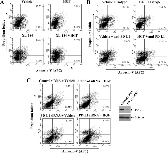
FIGURE 5.
c-Met-induced increase in PD-L1 protects renal cancer cells from immune cell-mediated cytotoxicity. A, RENCA cells/splenocytes co-culture (as described in “Experimental Procedures”) was pre-treated with either XL-184 (10 μm) or vehicle for 4 h. Following incubation, co-cultured cells were treated with either HGF (50 ng/ml) or vehicle. Following 72 h of treatment, the adherent RENCA cells were harvested and apoptotic index of the cells was analyzed by annexin V and propidium iodide staining. B, RENCA cells/splenocytes co-culture was incubated with either PD-L1 antibody (10 μg/ml) or isotype antibody for 8 h, and then treated with either HGF (50 ng/ml) or vehicle. Following 72 h of treatment, the adherent RENCA cells were harvested and apoptotic index of the cells was analyzed by annexin V and propidium iodide staining. C, RENCA cells were transfected with 50 nm of either PD-L1 siRNA or control siRNA. Following 48 h of transfection, cells were utilized in the RENCA cells/splenocytes co-culture and treated with either HGF (50 ng/ml) or vehicle. Following 48 h of treatment, the adherent RENCA cells were harvested and apoptotic index of the cells was analyzed by annexin V and propidium iodide staining. The Western blot (right panel) represents the siRNA-mediated knockdown of PD-L1 compared with β-actin control. A–C, representative data of three independent experiments.

