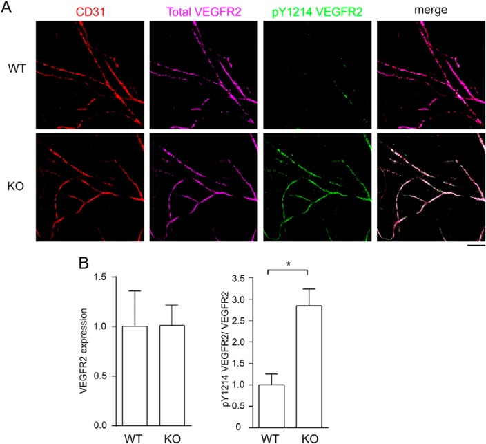
FIGURE 3.
Acute activation of VEGFR2 is elevated in dermal microvessels of R-Ras KO mice. A, VEGF-A (30 μl of 50 ng/ml) was injected intradermally into mouse ears, and the ears were collected 15 min later. The whole-mount immunofluorescence for CD31 (red), total VEGFR2 (purple), and Tyr(P)-1214 VEGFR2 (green) was imaged by confocal microscopy. Scale bar = 100 μm. B, quantitative analysis was carried out by Western blotting of the ear tissue lysate as described for the tumor studies. Four (WT) or six (KO) animals were examined per group. Error bars shown mean ± S.E. *, p < 0.05.

