
FIGURE 7.
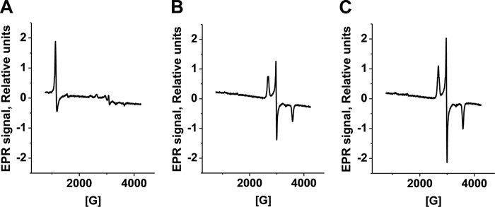
EPR spectra of MetHb treated with sulfide. Shown are EPR spectra of samples containing 50 μm MetHb in 100 mm HEPES, pH 7.4 (A), or after a 5-min incubation with 1200 μm Na2S under aerobic (B) or anaerobic (C) conditions at 25 °C. The EPR settings are described under “Experimental Procedures.”