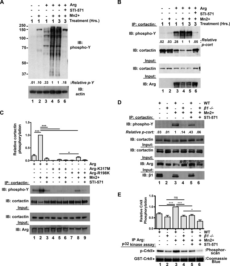
FIGURE 5.
Integrin activation potentiates Arg kinase activation in cells. A, integrin activation enhances Arg-dependent global protein phosphorylation in cells. Control or Arg-RFP-expressing Phoenix cells were treated with or without Mn2+ for 1 h to activate integrins. Some conditions were then lysed (lanes 1–4), whereas other conditions (lanes 5 and 6) were further treated with 5 μm STI-571 or DMSO vehicle control for an additional 2 h (3 h of total Mn2+ treatment) before cell lysis. Lysates were then separated on Bis-Tris PAGE gels and immunoblotted (IB) for phosphotyrosine. Integrin activation strongly enhances Arg-mediated phosphorylation of proteins in the lysate (compare lanes 3 and 4), but co-treatment of integrin activated Arg-RFP-expressing cells with 5 μm STI-571 (compared with DMSO vehicle control) attenuates this phosphorylation (compare lanes 5 and 6). B, integrin activation drives Arg-mediated cortactin phosphorylation. Control or Arg-RFP-expressing Phoenix cells were treated as in A, cortactin was immunoprecipitated (IP) from cell lysates and immunoblotted with anti-phosphotyrosine antibodies. Integrin activation results in elevated cortactin phosphorylation in Arg-RFP-expressing cells (compare lanes 3 and 4), which is attenuated by STI-571 treatment (compare lanes 5 and 6). C, the Arg SH2 domain is required for optimal integrin activation-mediated Arg kinase activation. Arg-YFP, kinase inactive Arg-K317M-YFP, and SH2 mutant Arg-R198K-YFP were expressed in Phoenix cells, and integrin activation assays were performed as in B. Arg-R198K-expressing cells exhibited intermediate levels of Arg-mediated cortactin phosphorylation, compared with WT and kinase inactive Arg-K317M-expressing cell conditions. Error bars represent S.E. from n = 3 for each condition. *, p < 0.05; ***, p < 0.001. D, integrin β1 activation drives Arg kinase activation. Isogenic WT or integrin β1−/− 3T3 fibroblast lines were treated with or without 2 mm Mn2+ for 2 h before lysing and immunoprecipitating cortactin. Immunoprecipitated products were then run on Bis-Tris gels, transferred, and immunoblotted for phosphotyrosine. Phosphorylated cortactin levels are markedly lower in integrin β1−/− 3T3 cells compared with WT, and Mn2+ induces a robust increase in phosphorylated cortactin in WT but not integrin β1−/− 3T3 cells. Image shown is representative of >3 independent experiments. E, Arg was immunoprecipitated from WT and integrin β1−/− 3T3 cells following treatments as performed in D, and its relative activity was measured in in vitro kinase assays using CrkII as a substrate. Mn2+-mediated integrin activation enhances Arg activity in WT, but not integrin β1−/− cells. Overnight STI-571 (20 μm) pretreatment dampens Arg activity via both readouts described in D and E. Error bars represent S.E. from n = 4. *, p < 0.05; ***, p < 0.001; ns, not significant.

