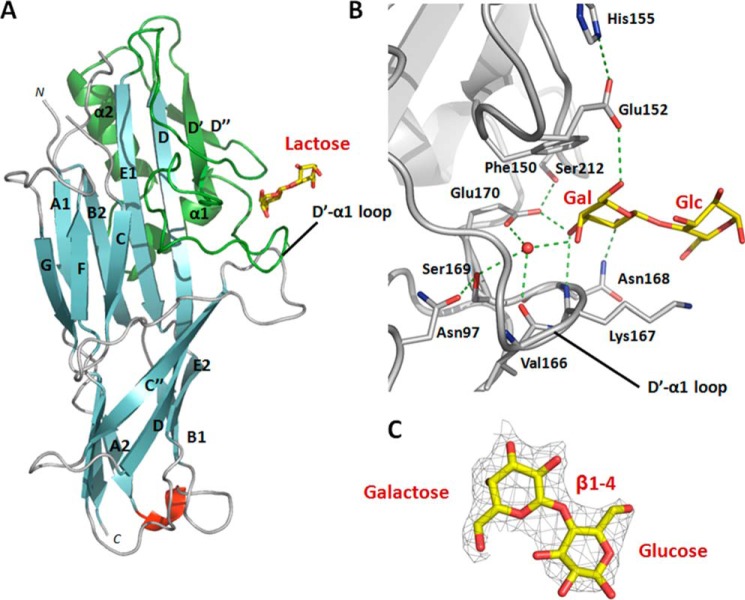
FIGURE 1.
Co-complex structure of the adhesive major subunit FaeGntd/dsc variant ad with lactose. A, graphic representation of FaeGntd/dsc variant ad bound to lactose (depicted in stick model). The lactose ligand is bound on the side of the FaeG major subunit by the D′-D″-α1-α2 subdomain (colored in green) grafted on the Ig-like core between strands D and E1. In particular, residues located on the D′-α1 loop are involved in complex formation. Strands are colored cyan, loops are in gray, and a short unassigned α-helix is in red. B, close-up of the FaeGntd/dsc variant ad binding site in complex with lactose. Two short amino acid stretches of the D′-α1 loop are involved in receptor binding. Only the galactose monosaccharide of the lactose ligand is interacting with binding site residues. Both side chains and main chain groups of residues on both stretches are coordinating a stabilizing water molecule (depicted as a red sphere). The protein backbone is in ribbon representation (gray), hydrogen bonds are depicted as dashed green lines, and ligand residues are displayed in stick representation with carbon and oxygen atoms in yellow and red, respectively. C, electron density map contoured at 1.3σ and displayed around the lactose ligand (depicted in stick model). Carbon and oxygen atoms are colored in yellow and red, respectively.

