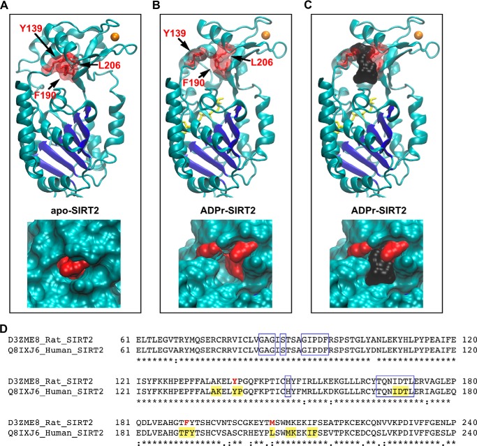
FIGURE 3.
A, top, the positions of residues that were photolabeled by AziPm are shown in the apo-SIRT2 structure. The Rossmann fold of SIRT2 is colored dark blue, zinc is colored orange, and the indicated photolabeled residues are shown as red sticks outlined by a transparent surface topology. Bottom, enlarged view of the photolabeled residues on the apo-SIRT2 structure, but with the solid surface topology of the protein shown. B, identical views as in A, but with the structure of ADPr-SIRT2. ADP-ribose is colored yellow. C, the cavity in the ADPr-SIRT2 structure is shown with a black surface representation, which has a volume of 364 Å3. D, segments of the human and rat SIRT2 sequences, which were derived from the indicated UniProt codes, are aligned. Identical residues are indicated by the asterisks, residues photolabeled by AziPm are bolded and red, residues lining the anesthetic cavity in ADPr-SIRT2 are highlighted yellow, and boxed residues form the protein C-pocket.

