
FIGURE 3.
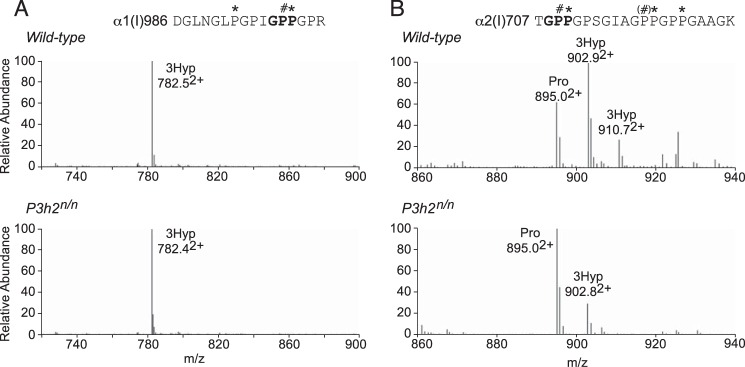
P3h1 substrate sites in type I collagen from mouse eye scleral tissue. LC-MS profiles of in-gel trypsin digests of the type I collagen α-chains from wild-type and P3h2n/n mouse sclera. A, MS profile of the α1-chain from the P3h2n/n mouse confirms no effect on Pro-986 3-hydroxylation (782.52+). B, MS spectrum from the α2-chain shows significant reduction of 3-hydroxylation at Pro-707 (895.02+) in the P3h2n/n mouse. A similar phenomenon was observed in type I collagen from cornea and tendon (data not shown). The trypsin-digested peptide is shown with P# indicating 3Hyp and P* indicating 4Hyp.