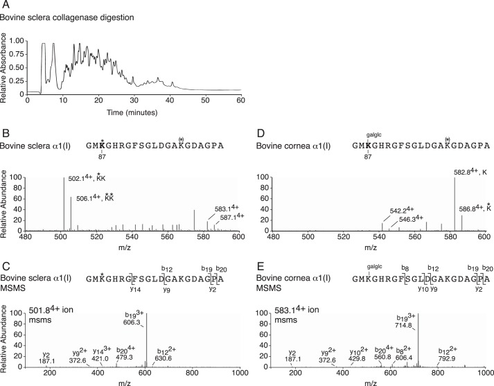
FIGURE 8.
Post-translational variances in cross-linking lysines between cornea and sclera type I collagen. A, profile of collagenase-digested whole tissue separated on C8 column (bovine sclera shown). B, LC-MS profile of C8 fraction 28 from bovine sclera reveals no glycosylation at cross-linking α1(I) Lys-87 (502.14+). C, MS/MS fragmentation spectrum of the parent ion (501.84+) from bovine sclera. The b and y ion breakages confirm no glycosylation on α1(I) Hyl87. D, LC-MS profile of C8 fraction 28 from bovine cornea reveals complete glycosylation of cross-linking α1(I) Hyl87 as glucosyl-galactosyl (galglc) (582.84+). E, MS/MS fragmentation spectrum of the parent ion (583.14+) from bovine cornea. The b and y ion breakages reveal gain of 340 Da (glucosyl-galactosyl) on α1(I) Hyl87. The trypsin-digested peptide is shown with K* indicating Hyl, K(*) indicating partial Hyl and galglc indicating glucosyl-galactosyl.

