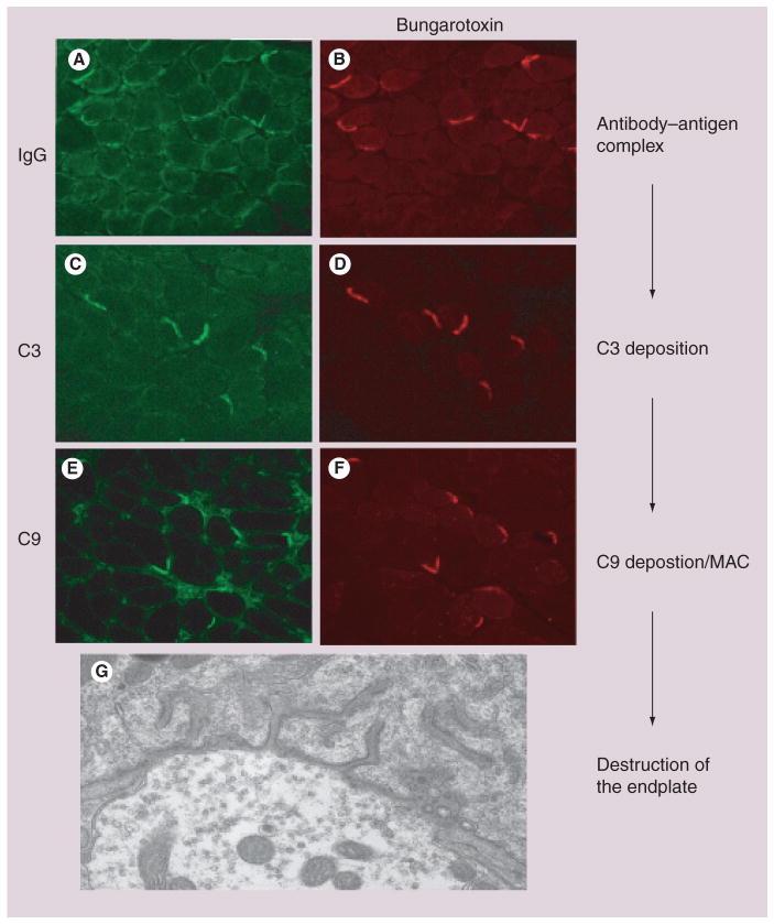
Figure 2. Complement deposition at the neuromuscular junction.
(A) Complement at the endplate begins with the binding of the antibody to the antigen on the cell surface. (B) Demonstrates the binding of IgG to the cell surface at the endplate that is marked by Texas red-bungarotoxin. (C & D) The initiation of the complement cascade occurs and allows for C3 to localize to the endplate. (E & F) The end product for the complement cascade is the deposition of the MAC, as demonstrated by C9 staining. (G) The hallmark feature of myasthenia gravis is the destruction of the endplate as shown as at the level of electron microscopy.
MAC: Membrane attack complex.

