
Figure 2.
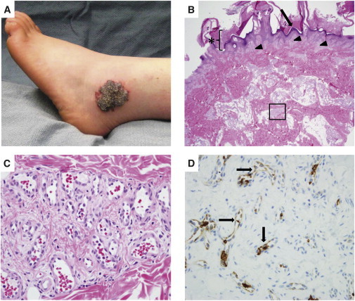
Photograph and Photomicrographs of the Verrucous Venous Malformation from Participant 5
(A) VVM lesion that contains the somatic mosaic MAP3K3 missense mutation.
(B) Hematoxylin-and-eosin-stained section of the excised VVM. Note hyperkeratosis (asterisk), papillomatous epidermis (arrow), and vascular clusters (arrowheads and boxed area) within the papillary dermis and reticular dermis, respectively, that extend into subcutaneous fat.
(C) Higher-magnification photomicrograph of the boxed area in (B) showing a cluster of venule-like channels.
(D) Immunostaining revealing GLUT1 expression in many endothelial cells within the VVM lesion (arrows).