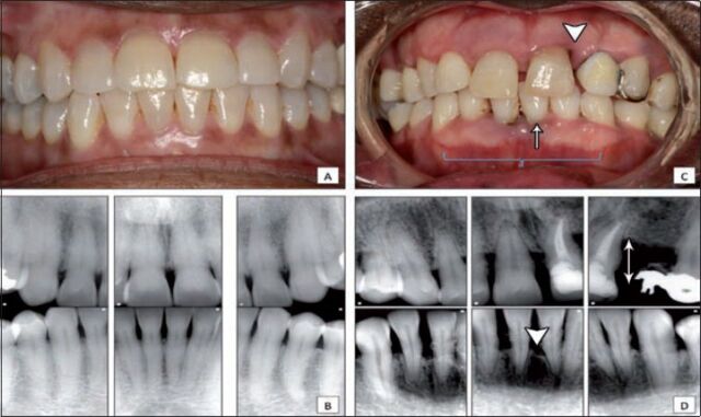
Figure 1.
Clinical comparison between healthy and periodontitis patients A) frontal intra-oral view for a patient with gingival health and stable periodontium demonstrating normal gingival color, marginal gingival level and intact inter-dental papillae; B) periapical radiographs for the patient in A, showing stable periodontium, normal bone level and absence of vertical bone defect; C) frontal intra-oral view of a patient with moderate to severe periodontitis presenting as loss of attachment (triangle), recession (arrow) and gingival edema (brace); D) periapical radiographs for the patient in C, showing calculus accumulation, horizontal bone loss (arrow) and vertical bone defect (triangle).

