Figure 3.
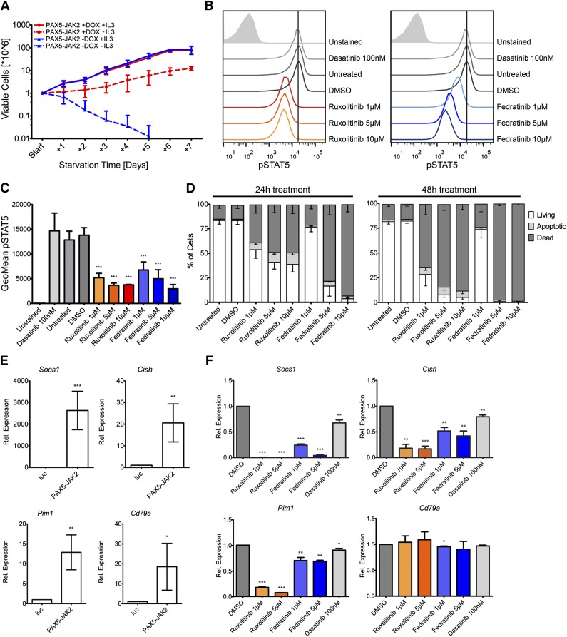
PAX5-JAK2 transforms Ba/F3 cells, and its kinase activity can be blocked by JAK2 inhibitors. (A) Growth rates of Ba/F3 cells in the presence (red) or absence (blue) of doxycycline with (full lines) or without (dashed lines) IL3 were monitored over 7 days by trypan blue exclusion staining. The means ± SD of 3 independent biological replicates are shown. (B) IL3-independent Ba/F3 cells expressing PAX5-JAK2 were treated with the indicated inhibitors for 1 hour. Intracellular pSTAT5 levels were measured by flow cytometry analysis. One of 3 representative experiments is shown. (C) Summary of geometric means ± SD (n = 3) of pSTAT5 levels as determined in B. (D) Cytokine-independent Ba/F3 cells expressing PAX5-JAK2 were treated with the indicated inhibitors for (left) 24 and (right) 48 hours. Staining for apoptotic/dead cells was performed using Annexin-V-allophycocyanin and DAPI, respectively. Means of 3 independent biological replicates are shown. Error bars represent SD. (E) Ba/F3 cells harboring the luc-control vector or PAX5-JAK2 vector were treated with doxycycline for 24 hours. Gene expression was measured using quantitative real-time PCR. Expression values were normalized using Abl1 and Gusb as control genes. Expression relative to luc-control of ≥3 independent experiments is shown. Significance levels were determined using an unpaired 2-tailed Student t test comparing the mean fold activation values of 3 independent experiments with the hypothetical value of 1 (*P < .05, **P < .01, ***P < .001). (F) Cytokine-independent Ba/F3 cells expressing PAX5-JAK2 were treated with indicated inhibitors for 8 hours. Relative gene expression was determined as in E.

