
Figure 1.
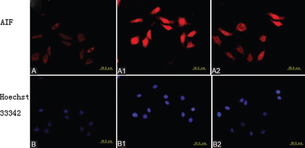
Immunofluorescent staining in each group (apoptosis inducing factor [AIF] nuclear translocation). (A) Cytoplasm emits red fluorescence in normal control group, AIF expression is mainly in cytoplasm and nuclei are olistherozone. (A1) Both cytoplasm and cell nuclei emit red fluorescence, but AIF expression is mainly in cell nuclei in oxygen and glucose-deprived group, and red fluorescence in the cytoplasm is weaker than that in normal control group, demonstrating that AIF nuclear translocation initiates ap-optosis. (A2) Both cytoplasm and cell nuclei emit red fluorescence in extract of Ginkgo biloba-pretreated group, but the red fluorescence in the nuclei is weaker than that in oxygen and glucose-deprived group. (B) Cell nuclei emit dispersed blue fluorescence after Hoechst 33342 staining in normal control group. (B1) Cell nuclei emit blue florescence after Hoechst 33342 staining, and the blue fluorescence is stronger in oxygen and glucose-deprived group than in normal control and Extract of Ginkgo biloba-pretreated groups. (B2) Cell nuclei emit blue fluorescence after Hoechst 33342 staining in extract of Ginkgo biloba-pretreated group, and the blue fluorescence in partial cell nuclei is weaker than that in oxygen and glucose-deprived group (AIF: Apoptosis inducing factor; OGD: Oxygen and glucose-deprived; EGb761: Extract of Ginkgo Biloba)