
Figure 2.
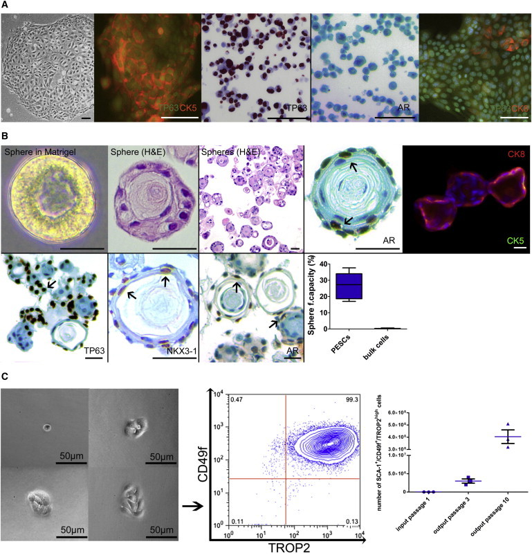
Characterization and Differentiation of Murine Basal PESCs
(A) IHC and immunofluorescence characterization of 2D cultured murine basal PESCs. Scale bar, 100 μm.
(B) Characterization of differentiated murine prostaspheres. Morphology in semisolid Matrigel; immunofluorescence and IHC of prostaspheres. Scale bar, 100 μm. For internal validation of TP63/AR/NKX3-1 antibodies, see Supplemental Experimental Procedures. The sphere-forming capacity of enriched PESCs was compared with bulk digested cells, n = 5 independent PESC preparations, p < 0.01 as determined by Student’s two-tailed t test.
(C) In vitro self-renewal. Colonies derived from single-cell-sorted cultured PESCs retain their SCA-1+/CD49f+/TROP2high phenotype after colony outgrowth. Amplification (cell numbers) of SCA-1+/CD49f+/TROP2high cells using the MPM culture method, n = 3.