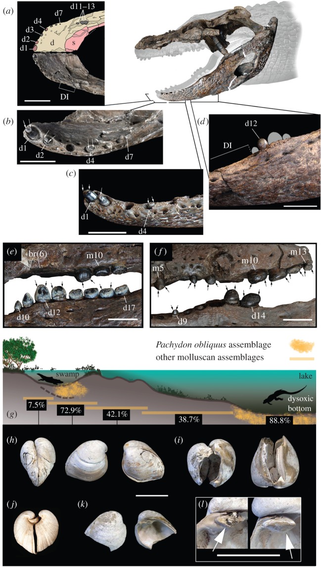
Figure 3.
Crushing-dentition caimanines and co-occurring pachydontine molluscs. (a–d) Gnatusuchus pebasensis. MUSM 662, anterior mandibular anatomy in dorsal (a), anterodorsal (b) and lateral view (c). (d) MUSM 2040, posterior mandibular dentition in lateral view. (e) Kuttanacaiman iquitosensis, MUSM 1942, posterior dentition. (f) Caiman wannlangstoni, MUSM 1983, posterior dentition. (g) Inferred distribution of molluscan assemblages within the typical depositional environments of the Pebas System [2]. Percentage values correspond to the estimated average abundance of the Pachydon group within each assemblage. Thick-shelled Pachydon obliquus (h,i,k,l) and Pachydon cuneatus (j) with convex outline making them well capable to withstand external pressure. (h,i) Crushing type predation scars in specimens that survived and then resumed growth. (k) Sharp edges typical of this type of predation. (l) Detail of cardinal tooth with (left) and without (right) crushing damage. See electronic supplementary material, and [2] and [4] for molluscan data and localities. Arrows in (b,c,e,f) indicate severe crown tooth wear. d, dentary; d1–4, d7, d9–14, d17, dentary tooth positions; DI, diastema; br(6), bone resorption in alveolar position 6; m5, m10, m13, maxillary tooth positions; s, splenial. Scale bars: 5 cm for (a), 2 cm for (b–f) and 1 cm for (h–l).

