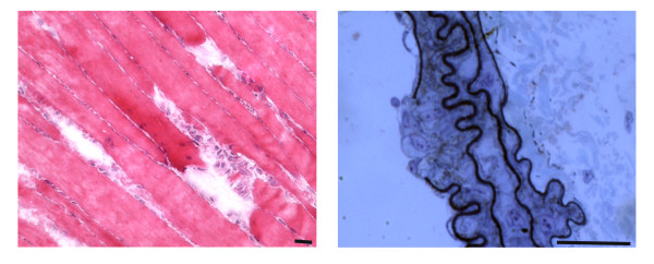
Figure 1.

Examples of focal injuries. (LEFT) Hematoxilin-and eosin-stained murine skeletal muscle, longitudinally sectioned to show the gaps in three adjacent fibers. The injury likely occurred following an intense exercise session (wheel running). Upon leakage of the broken sarcolemma, factors such as Wnt are released before a fast repair process known as patch repair occurs. In turn, Wnt factors trigger the activation of satellite cells and other resident interstitial cells with myogenic potential, which proliferate, migrate and fuse into small myotubes that ultimately fuse with the damaged fibers. (RIGHT) Toluidine blue-stained semithin section of a murine carotid showing damage, likely due to smooth muscle cell-restricted inactivation of the serum response factor gene. A rupture of the endothelial layer, as well as of the elastin matrix, with exposure of underlying cells is visible; release of intracellular factors (von Willebrand Factor) and exposure of undisclosed antigens (collagen) are essential for the subsequent phases of clot formation, remodeling and repair of the wall defect. Bar = 25 micron.
