
Figure 3.
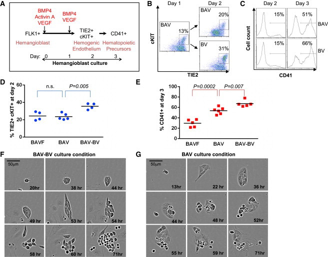
Activin A Impairs the Maintenance of HE
(A) Schematic representation of the experimental strategy. FLK1+ cells sorted from day 3 EBs were seeded on gelatinized plates in serum-free media supplemented with BAV for the first day and then with BV from day 1 onward (B, BMP4; A, Activin A; V, VEGF).
(B) Flow cytometry analysis of TIE2 and cKIT coexpression at day 1 and 2 of FLK1+ cell culture grown in a BAV or BAV-BV cytokine combination.
(C) Flow cytometry analysis of CD41 expression at days 2 and 3 of the same cultures.
(D and E) Graph of data obtained from BAFV, BAV, and BAV-BV cultures, showing the frequencies of TIE2+cKIT+ cells at day 2 (D) and CD41+ cells at day 3 (E). Each point represents an independent experiment.
(F and G) Representative time-lapse imaging of FLK1+ sorted cells cultured in serum-free media supplemented with a BAV-BV (F) or BAV (G) cytokine combination. Data shown are representative of at least three independent experiments (n.s., nonsignificant).
See also Figure S2.