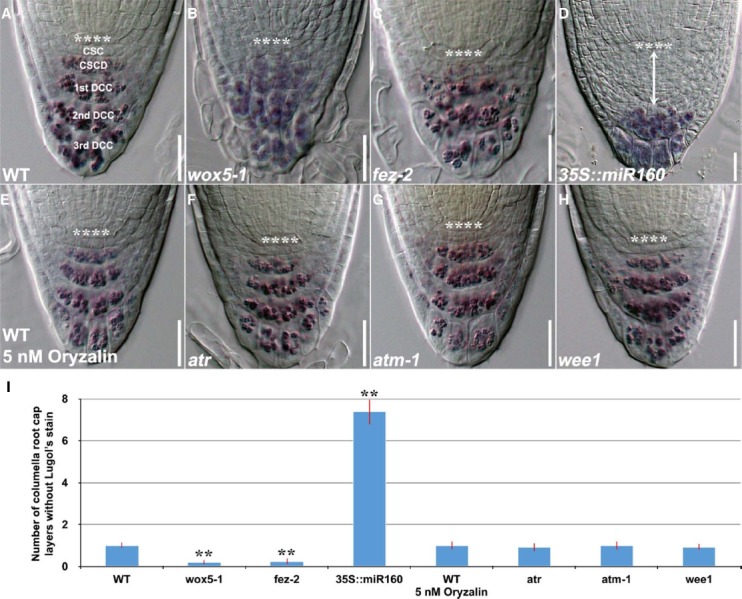
FIGURE 1.

Lugol’s staining showing accumulation patterns of starch granules in the columella root cap of 4-day-old WT and mutant genotypes. (A) WT (n = 14). (B) wox5-1 (n = 15). (C) fez-2 (n = 11). (D) 35S::miR160 (n = 15). (E) WT treated with 5 nM oryzalin for 24 h (n = 11). (F) atr (n = 12). (G) atm-1 (n = 12) and (H) wee1 (n = 12). (I) Quantification of the number of layers of unstained columella root cap cells. Error bars represent standard error of the mean. **: P < 0.01, t-test. **** represents position of the QC. CSC: layer of columella root cap stem cells; CSCD: layer of differentiating columella root cap stem cell daughters; 1st DCC: 1st layer of fully differentiated columella root cap cells; 2nd DCC: 2nd layer of fully differentiated columella root cap cells; 3rd DCC: 3rd layer of fully differentiated columella root cap cells. Scale bars: 20 μm.
