FIGURE 3.

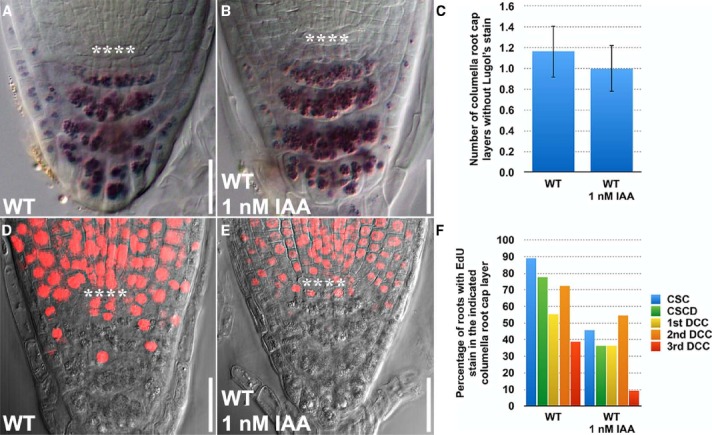
EdU and Lugol’s staining can be combined and jointly used to investigate the effects of exogenously applied auxin on stem cell homeostasis in the Arabidopsis root. (A) Lugol’s staining of WT (n = 14). (B) Lugol’s staining of WT treated with 1 nM IAA for 24 h (n = 13). (C) Quantification of the number of layers of unstained columella root cap cells. (D) EdU staining of WT treated with 10 μM EdU for 24 h (n = 18). (E) EdU staining of WT treated with 10 μM EdU and 1 nM IAA for 24 h (n = 13). (F) Quantification of the percentage of roots with EdU stain in the indicated columella root cap layer. Error bars represent standard error of the mean. **** represents position of the QC. CSC: layer of columella root cap stem cells; CSCD: layer of differentiating columella root cap stem cell daughters; 1st DCC: 1st layer of fully differentiated columella root cap cells; 2nd DCC: 2nd layer of fully differentiated columella root cap cells; 3rd DCC: 3rd layer of fully differentiated columella root cap cells. Scale bars: 20 μm.
