FIGURE 3.
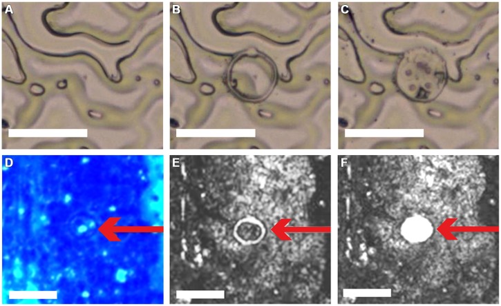
Dissection and catapulting of 30 μm-disks from ACT-prepared adaxial leaf epidermis. Application of LM coupled with laser pressure catapulting (LMPC) on ACT-prepared A. thaliana adaxial leaf epidermis. Epidermis (A) before and (B) after microdissection of a 30 μm disk; (C) removal of 30 μm-disk from epidermal tissue by LMPC. (D) ACT-prepared adaxial leaf epidermis 3 days post-inoculation with the powdery mildew Golovinomyces cichoracearum, additionally stained with aniline blue to visualize callose deposition. Red arrow indicates 30 μm-disk including pathogen-induced, callosic papillae (spots with light-blue fluorescence). (E,F) Bright-field micrographs of the same epidermis area as in (D,E) after microdissection and (F) after LMPC of a 30 μm-disk. PALM MicroBeam LM system used for LMPC and micrograph acquisition. Scale bars = 50 μm.

