Figure 6.
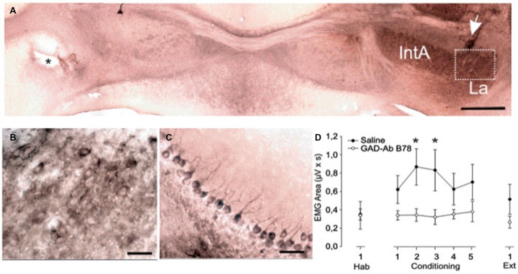
Learning curves and eyelid performance collected from controls (saline) and experimental (b78) mice during a delay conditioning paradigms. Spreading of b78 in the deep cerebellar nuclei was confirmed by immunohistochemistry. (A) Presence of b78 in deep nuclei areas of the right cerebellar hemisphere. The injection site in the contralateral area is indicated by the asterisk. (B) Enlargement of the marked area in panel (A). (C) Purkinje cells of a lobule area proximal to the injection site. Abbreviations: IntA, anterior part, La, lateral part. Calibration bars: (A): 200 μm, (B,C) 50 μm. (D) Conditioned eyelid responses evoked in controls were significantly larger (F(1,14) = 4.6; P = 0.05; see asterisks) than those evoked in b78 animals.

