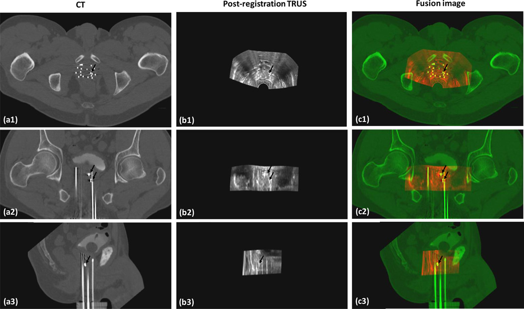
Fig. 4. TRUS-CT registration results.
a1-a3 are CT images of a 58-year-old prostate-cancer patient; b1-b3 are his post-registration TRUS images; and c1-c3 are the fusion images between the CT and post-registration TRUS images. The close match between the gold markers (dark arrows) on the CT and TRUS demonstrated the accuracy of our registration method.

