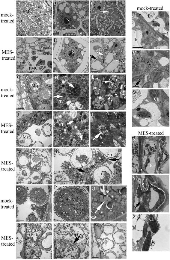
Figure 1.

Transmission Electron Microscope (TEM) micrographs of the anthers from the mock-treated (fertile) and MES-treated (sterile) plants. (A) The fertile anthers at pollen mother cell (PMC) stage; (B) Enlarged fertile meiocytes in (A); and (C) Enlarged fertile tapetum in (A) showing numerous plastids dispersed in cytoplasm (white arrow). (D) The sterile anthers at PMC stage; (E) Enlarged sterile meiocytes in (D) showing less plastids in condensed cytoplasm separated from the cell wall; (F) Enlarged sterile tapetum in (D) showing little abnormal plastids (white arrow) and more large vacuoles in cytoplasm, and with a little plasmolysis at meiocyte side (black arrow). (G) The fertile anthers at vacuolated-microspore stage; (H) The degraded tapetum in (G) showing elaioplasts and tapetsomes with abundant lipids; (I) Plastids in tapetum located in a crown showing filled with globular low electron-dense metabolites and surrounded by rich endoplasmic reticulum (ER). (J) The sterile anthers at vacuolated-microspore stage (type I); (K) The undegraded tapetum in (J) showing elaioplasts and tapetsomes with abundant lipids; (L) Plastids in tapetum located in a crown showing irregular shaped low electron-dense material. (M) The sterile anthers at vacuolated-microspore stage (type II); (N) The degraded tapetum in (M) showing scattered elaioplasts and tapetsomes with fuzzy structure; (O) The fertile anthers at mature pollen grain stage; (P) The pollen grain in (O) showing profuse globular particles; (Q) The enlarged globular particles in (P). (R) The sterile anthers at mature pollen grain stage (type II); (S) The undegraded tapetum in (R) died but cell wall still existed (black arrow); (T) The sterile anthers at mature pollen grain stage (type I). (U) The epidermis and endothecium cells in fertile plants at vacuolated-microspore stage; (V) The epidermis cells in (U) showing normal oval-shaped chloroplastids with distinct thylakoid structure and little starch granules in thylakoid; (W) The endothecium cells in (U) showing oval-shaped chloroplastids with distinct thylakoid structure. (X) The epidermis and endothecium cells in sterile plants at vacuolated-microspore stage; (Y) The epidermis cells in (X) showing abnormal chloroplastids with large starch granules in thylakoid; (Z) The endothecium cells in (X) showing fusiform-shaped chloroplastids with linear thylakoid structure. PMC, pollen mother cell; N, nucleus; T, tapetum; Msp, microspore; Ep, elaioplast; Ts, tapetosome; PG, pollen grain; TCW, tapetum cell wall; E, epidermis; En, endothecium; Ch, chloroplast. Scale bars = 10 μm (A, D, G, J, M, N, O and T), 5 μm (C, F, P, R, S, U and X), 2 μm (B, E and K), and 1 μm (H, I, L, Q, V, W, Y and Z).
