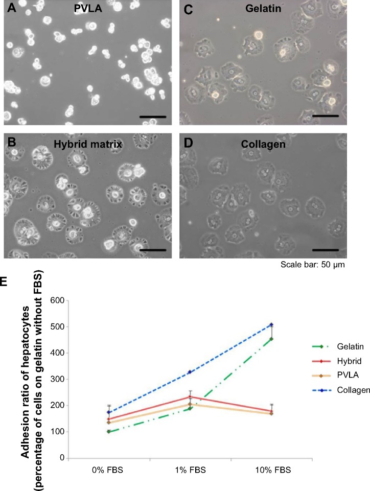
Figure 2.
The attachment of primary hepatocytes in various systems (4 hours).
Notes: The cell morphology on PVLA with 1% FBS (A), the hybrid matrix with 1% FBS (B), gelatin with 10% FBS (C), and collagen with 10% FBS (D) is shown. The relative amount of attached hepatocytes was further determined by using the number of cells on the gelatin surface without FBS as the 100% value (E). The cell numbers on gelatin and on collagen increased when the serum concentration ranged from 0% to 10%, and they reached the highest level on the PVLA-containing matrices in the medium with 1% FBS. The hybrid matrix was made up of EFC and PVLA. Scale bar: 50 μm.
Abbreviations: FBS, fetal bovine serum; PVLA, poly-(N-p-vinylbenzyl-4-O-β-D-galactopyranosyl-D-gluconamide); EFC, E-cadherin-Fc.

