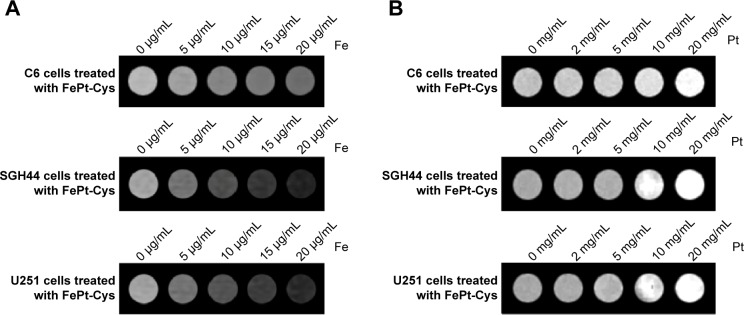
Figure 8.

Imaging of different gliomas cells (C6, SGH44, and U251) treated with FePt-Cys NPs.
Notes: (A) MR imaging of different gliomas cells at different Fe concentrations; (B) CT imaging of different gliomas cells at different Pt concentrations.
Abbreviations: CT, computed tomography; FePt-Cys, l-cysteine coated FePt; MR, magnetic resonance; NPs, nanoparticles.
