
Fig. 2.
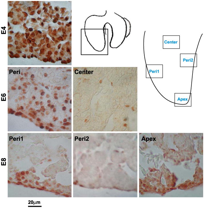
Identification of the LoGZ during liver development. Immunohistochemistry of PCNA staining at E4, E6, and E8. PCNA is all over in early liver stages but becomes restricted to several localized growth zones in the periphery and apex at later stages. Schematic drawings show the locations of the panels taken. Peri (periphery) 1 represents localized growth zones. Peri2 represents regions without growth zone activity. Scale bars, 20 μm.