Fig. 3.
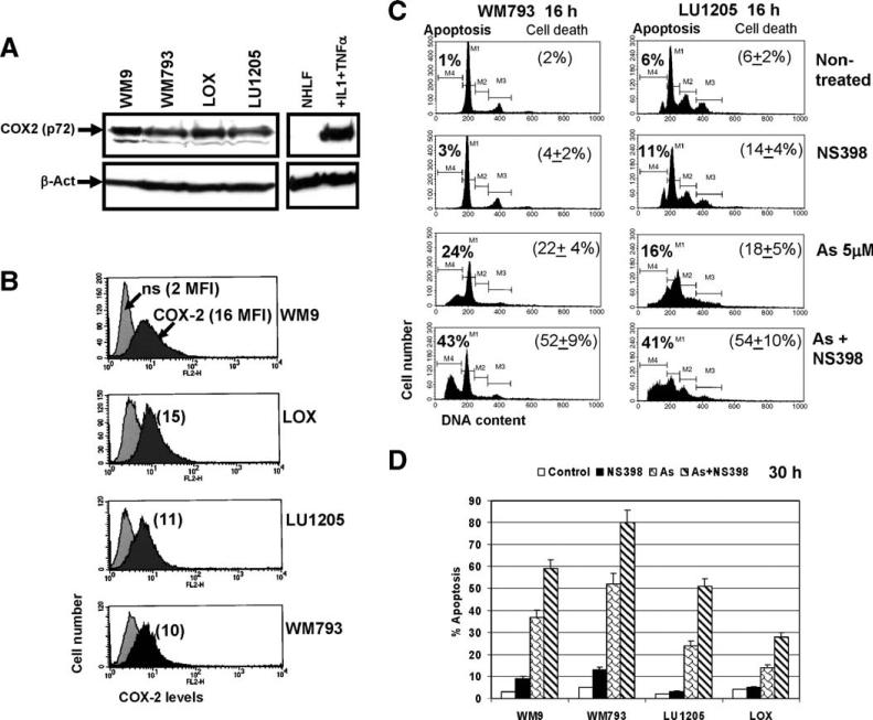
COX-2 expression in human melanomas. (A) Western blot analysis of COX-2 levels in the indicated cell lines. β-actin was used as a loading control. Normal human lung fibroblasts were untreated or treated with IL-1 (2 ng/ml) and TNFα (10 ng/ml) for 4 h. (B) Total COX-2 levels in human melanomas have been determined by FACS analysis after cell permeabilization and staining using mAb against COX-2, PE-conjugated goat anti-mouse secondary Ab and the flow cytometry. Nonspecific (ns) staining; medium fluorescence intensity (MFI) is indicated in brackets. (C, D) Inhibition of COX-2 activity by NS398 (50 μM) had synergistic effects on arsenite-induced apoptosis in COX-2-positive human melanomas. Levels of apoptosis have been determined by FACS analysis of PI-stained melanoma cells 16 h (C) and 30 h (D) after treatment with sodium arsenite (5 μM), NS398 (50 μM) or their combination; levels of total cell death have been determined by Trypan blue staining. Error bars represent mean ± SD from three independent experiments.

