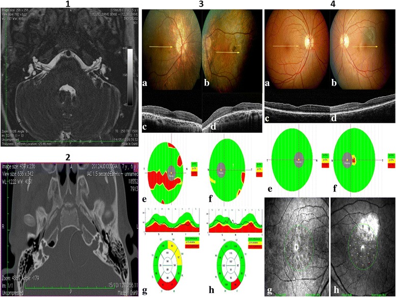
Figure 2.

Images for subject IV-1: 1) Axial MR TSE drive T2-weighted image. Note the small bulbous dilatation of the lateral part of the IAC, with absence of the modiolus and of the bone at the fundus. The cochlea is dysmorphic and small in size. The vestibule is dilated bilaterally. Note also the dilatation of the vestibular acqueduct on the right side. 2) Axial CT. The absence of the bone fundi of IAC and of the modiolus are better depicted on CT. Ophthalmic results: 3) Male patient affected with choroideremia. Color fundus images (a, right eye; b, left eye) and cross sectional OCT B-scan (c, right eye; d, left eye) show diffuse atrophy of the retinal pigment epithelium and choriocapillaries and round retinal pigment changes at the posterior pole and midperiphery. Ganglion cell complex thickness shows a severe arciform thinning in (e) the right eye, while focal alterations are present in (f) the left eye. Retinal nerve fiber layer is reduced in the inferior quadrants in (g) the right and (h) left eye. 4) Female patient carrier of the choroideremia gene. Color fundus images (a, right eye; b, left eye), cross sectional OCT B-scan (c, right eye; d, left eye), ganglion cell complex thickness map (e, right eye; f, left eye) and microperimetry (g, right eye; h, left eye) are normal. A choroidal nevus is visible at the posterior pole in the left eye.
