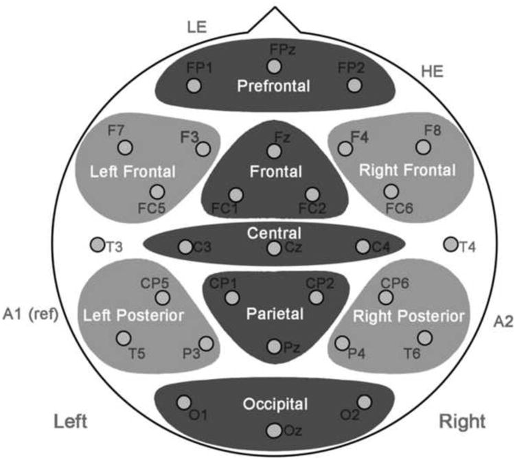
Figure 6.

Electrode montage, illustrating midline and peripheral regions for analysis of ERP data along with placement of left and right eye electrodes (LE, HE) as well as left mastoid reference (A1) and right mastoid for differential activity (A2).

Electrode montage, illustrating midline and peripheral regions for analysis of ERP data along with placement of left and right eye electrodes (LE, HE) as well as left mastoid reference (A1) and right mastoid for differential activity (A2).