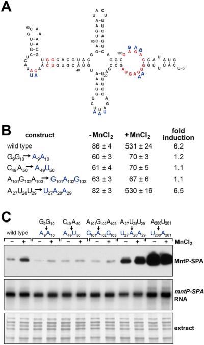
Figure 3. mntP riboswitch responds to MnCl2 in vivo and in vitro.
(A) Predicted secondary structure of the mntP aptamer based on conservation and structure probing. Nucleotides in red are conserved in >97% of the yybP-ykoY family members (Meyer et al., 2011). Blue letters correspond to mutants assayed in (B-C). See also Figure S1A and Figure S2.
(B) β-galactosidase activity for strains carrying PLlacO-5′UTRmntP-lacZ translational fusions with mutations of indicated residues grown in M9 glucose medium and incubated without and with 400 μM MnCl2 for 1 h. The results are given in Miller units as the mean ± SDM of three independent samples.
(C) Reconstitution of mntP responsiveness to manganese in vitro with a purified E. coli in vitro translation system. Wild type or mutant RNA (1 μg) encompassing the mntP 5′-UTR and mntP ORF with a C-terminal SPA tag was incubated in the presence of no metal or 400 μM MnCl2 for 2 h and then subjected to western blot analysis. The levels of the mntP-SPA mRNA were determined by primer extension analysis. The proteins were detected by silver staining of the gel after the transfer of the lower molecular weight proteins.

