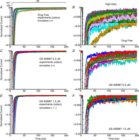
Figure 1.

Simulated and experimentally recorded INa data from isolated guinea pig ventricular myocytes
Model-generated INa in guinea pig ventricular myocytes (black) is shown superimposed on experimental INa records. The right panels show late INa at high gain. Currents were normalized to peak values. Normalized INa is shown in the absence of GS-458967 (A, n = 30 for experiments) and presence of 0.3 μm (B, n = 10) and 1 μm (C, n = 10) GS-458967.
