
Figure 9.
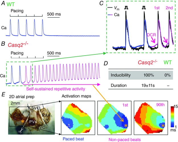
Optical mapping demonstrating repetitive focal activity in Casq2−/− atrial tissue preparations
A, representative Ca transient during and after pacing in WT atrial preparation. B, representative Ca transient during and after pacing in Casq2−/− atrial preparations. Pink colour indicates the presence of self-sustained repetitive activity. C, simultaneous optical membrane potential (Vm) and Ca signals during the initiation of self-sustained repetitive activity (marked by green dotted rectangles in B). Diastolic Ca releases (DCRs) are marked by the pink arrows. D, inducibility and duration of the repetitive activity under 100 nm isoproterenol in WT (n = 5) and Casq2−/− atrial tissue (n = 5). E, activation maps showing the sequence of electrical conduction from blue to red colours during pacing, 1st non-paced beat, and 90th non-paced beat during the recording in B. Bottom left is a representative unwrapped 2D atrial tissue preparation, where the left atrial appendage (LAA) is separated from the pacemaker complex and studied. RAA, right atrial appendage; SVC, superior vena cava; IVC, inferior vena cava; PVs, pulmonary veins.