
Figure 6.
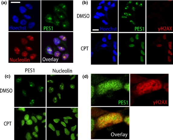
Endogenous PES1 localization and its redistribution after DNA damage. (a) Confocal images of TNB1 neuroblastoma cells immunostained for PES1 (green) and nucleolin (red) with nuclear stain (Hoechst33258, blue). PES1 is encircled by nucleolin, which shows “donut-like” staining. (b,c) Confocal images of TNB1 cells after DMSO or camptothecin (CPT; 1 μM) treatment. Scale bar = 15 μm. (d) Enlarged confocal images of TNB1 cells after CPT (1 μM) treatment. One nucleus was enlarged.