
Figure 1.
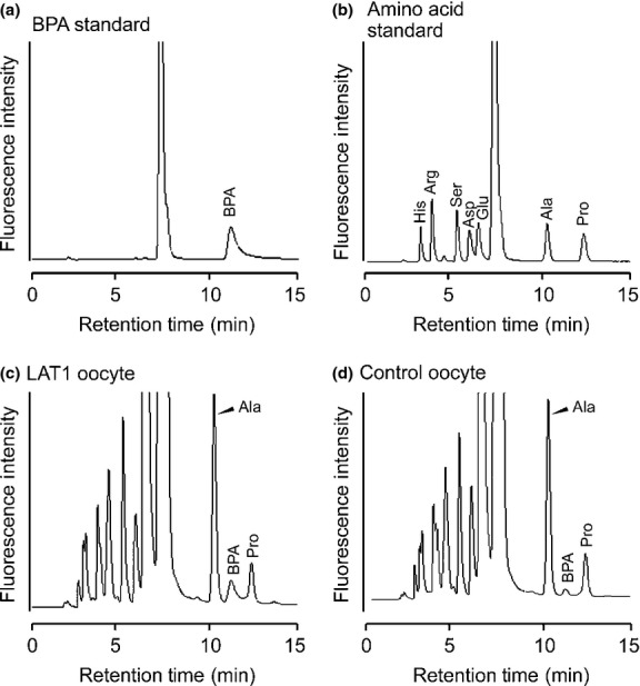
Separation of p-boronophenylalanine (BPA) by HPLC. Chromatograms showing the separation of a BPA standard (0.2 pmol) (a), amino acid standards (0.2 pmol each) (b), a sample from the oocyte expressing LAT1 (c), and the control oocyte not expressing LAT1 (d). (a, b) The BPA peak was identified by retention time and spike study (not shown). Similarly, by comparison with amino acid standards and a spike study (not shown), the peaks neighboring BPA in oocyte samples were identified as alanine (Ala) and proline (Pro). (c, d) The LAT1-expressing oocyte and non-expressing control oocyte were incubated in the uptake buffer containing BPA. Samples from the oocytes were separated by HPLC. The increased BPA peak height in (c) showed that the uptake of BPA was mediated by LAT1. Arg, arginine; Asp, aspartic acid; Glu, glutamic acid; His, histidine; Ser, serine.