
Figure 4.
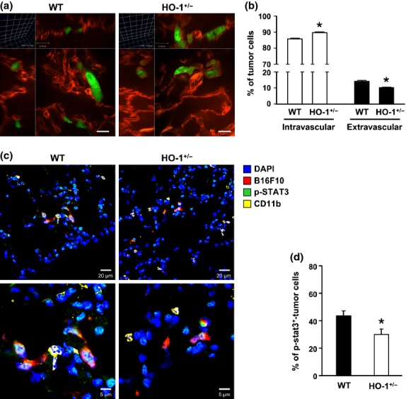
Heme oxygenase-1 (HO-1) influences extravasation and STAT3 activation of metastatic tumor cells. (a,b) Wild type (WT) and HO-1+/− mice (n = 5/group) received an i.v. injection of 1 × 10⁶ CMFDA-labeled B16F10 cells for 4 h. Pulmonary vasculature was labeled by isolectin IB4 Alexa Fluor 647 conjugates. (a) Representative 3-D confocal images of lungs. Bar = 12 μm. (b) The percentages of intravascular and extravascular tumor cells were determined. *P < 0.02 versus WT group. (c) WT and HO-1+/− mice received an i.v. injection of 1 × 10⁶ CMTMR-labeled B16F10 cells for 48 h. The lung sections were subjected to immunofluorescence staining with antibodies against p-STAT3 and CD11b, respectively. Data shown is the representative image. (d) The percentage of B16F10 cells with positive p-STAT3 immunostain in each group of mice (n = 3/group) was determined. *P < 0.02 versus WT group.