
Figure 4.
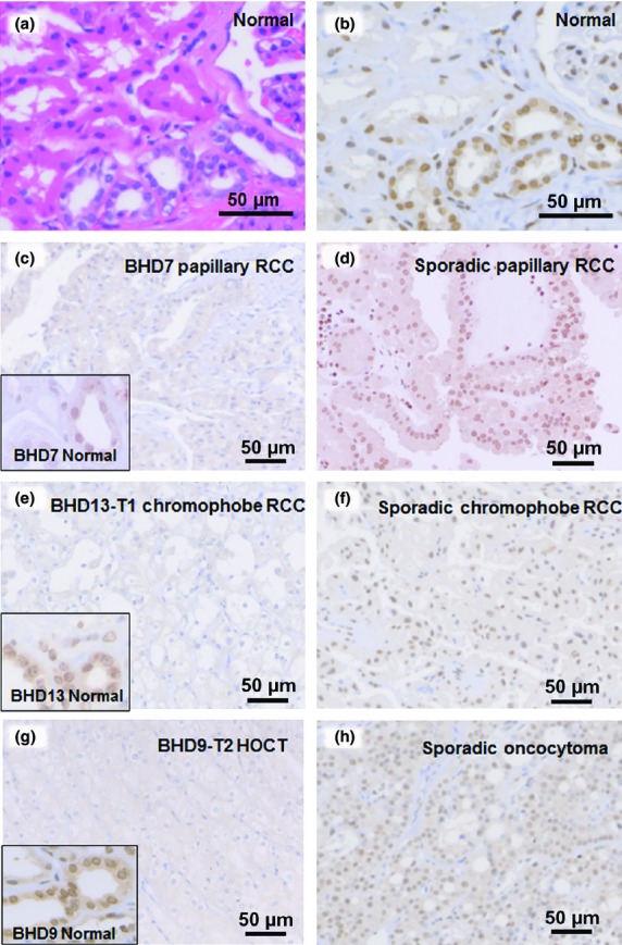
Folliculin (FLCN) immunostaining in normal kidney, and Birt–Hogg–Dubé (BHD) and sporadic tumors. (a) H&E staining of a normal kidney. (b) Serial section of sample A immunostained for FLCN. Nuclei of distal tubules were strongly positive for FLCN. (c–h) Immunostaining for FLCN in BHD and sporadic tumors. Weak cytoplasmic staining was evident in tumors of BHD patients (c, e, h). Insets highlight distal tubules of non-tumor areas, showing immunoreactivity to FLCN. Sporadic renal tumors (d, f, h) showed strong nuclear staining.