
Figure 5.
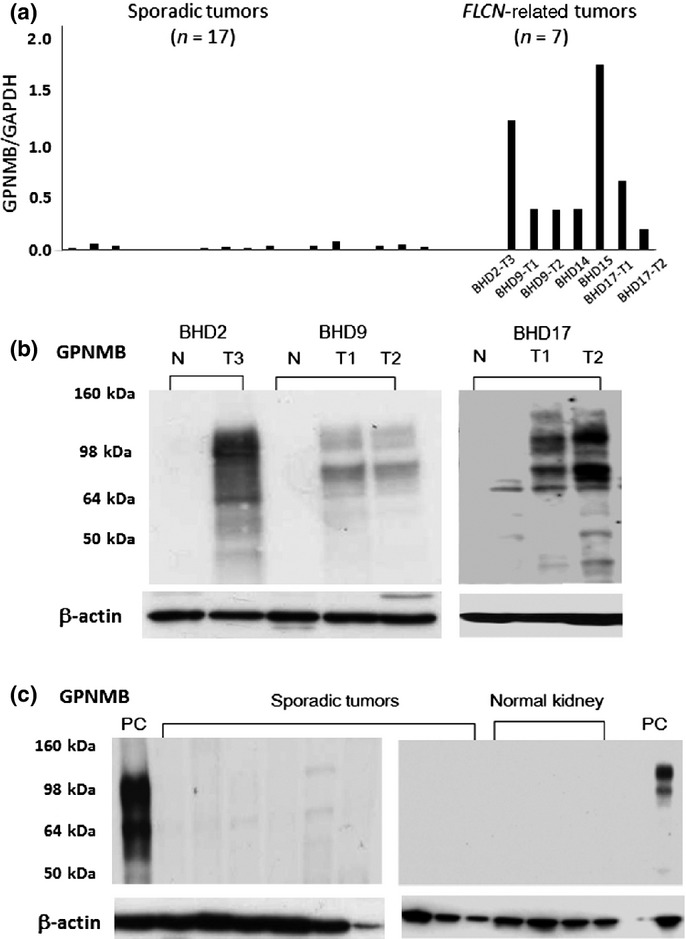
Glycoprotein non-metastatic B (GPNMB) expression in Birt–Hogg–Dubé (BHD) and sporadic tumors. (a) Expression levels of GPNMB mRNA were analyzed by quantitative RT-PCR as shown. Sporadic renal tumors include clear cell renal cell carcinomas (RCCs) (n = 9), oncocytoma (n = 1), papillary RCC (n = 1), and chromophobe RCCs (n = 6). (b) Representative results of Western blot analysis of BHD kidneys. Patients BHD9 and BHD17 had two independent tumors (T1, T2), respectively. Two isoforms (115 kDa and 80 kDa) of GPNMB bands were seen in tumor lanes, but not in normal-looking lanes. N, normal-looking region; T, tumor region. (c) Representative results of Western blot analysis of sporadic renal tumors and normal kidneys without the background of BHD. GPNMB bands were barely seen in sporadic tumor and normal kidney lanes. Sporadic renal tumors included clear cell RCCs (n = 3), oncocytoma (n = 1), papillary RCC (n = 1), and chromophobe RCCs (n = 4). PC, positive control using BHD9-T2.