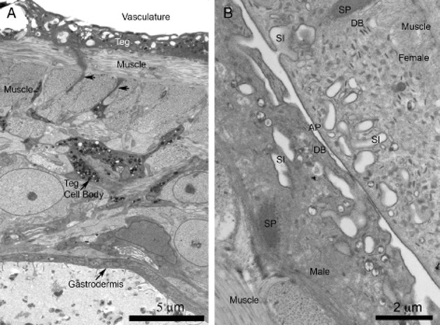
Figure 1.

Tegument of Schistosoma mansoni by transmission electron microscopy. (A) Low magnification image from a cross-section of an adult. The image shows the apical cytoplasm of the tegument (Teg), which is the interface with the host vasculature. The syncytial cytoplasm rests on bands of musculature and is supported and maintained by tegumentary cell bodies. That depicted is rich in vesicles that are transported to the apical cytoplasm along cytoplasmic bridges (arrows). The parasite digestive system, lined by a syncytial epidermis called a gastrodermis, lies deep within the body. (B) High magnification view of the teguments of paired male and female adults. The apical membrane complex (AP) consists of a plasma membrane overlain by a subsidiary membrane-like structure, the membranocalyx, evident only after special fixation/staining of TEM tissues using uranyl acetate. The apical cytoplasm infolds frequently as surface invaginations, sometimes with secondary caveola-like outpocketings appearing. Other bodies decorate the tegument, including discoid bodies (DB). Tegumentary spines (SP), used for adhesion, are observed. Original figures.
