
Figure 7.
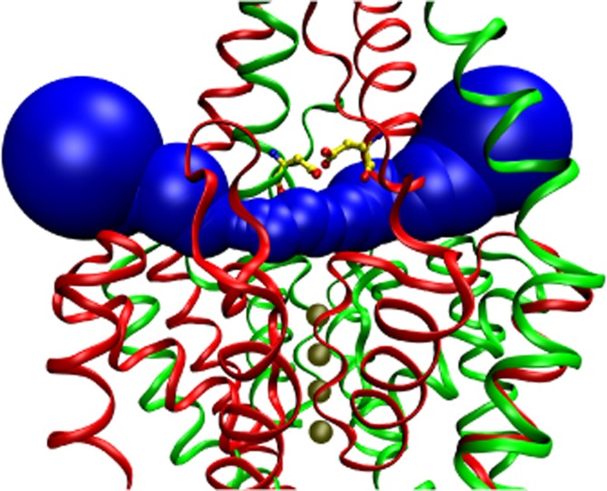
Aspartate 135 residues are positioned in the EIP above the selectivity filter. The EIP tunnel is filled with a series of blue spheres in this schematic representation of human TREK-2 crystal structure (PDB ID: 4BW5, A. C. W. Pike, et al., unpublished). K+ ions in the vertical channel pore are illustrated with four solid (tan) spheres below the EIP. Aspartate 135 (D135) residues (yellow ball and stick representation, with red oxygen and blue nitrogen atoms) are located on the ceiling of the EIP above the selectivity filter. The two subunits are drawn as red and green ribbons. The view is not exactly perpendicular to the direction of the EIP, but the structure is slightly rotated around the vertical axis, in order to avoid the overlap of the D135 residues of the two subunits. The NSSN and NSSNNS sequences of the subunits, respectively, are not resolved by the crystal structure; however, this does not affect the EIP. The graphics were produced with MolAxis (Yaffe et al., 2008) and VMD (Humphrey et al., 1996) software.