Figure 8.

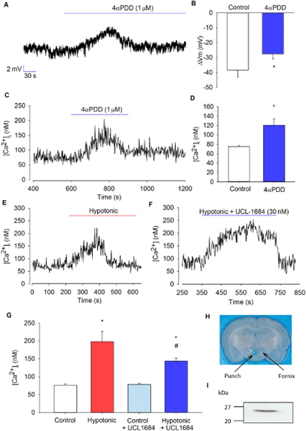
Whole-cell and Ca2+ in isolated PVN neurones. (A) Representative whole-cell current-clamp trace showing depolarization of the cell upon addition of 4αPDD. (B) Mean resting membrane potential of −54 ± 5 mV was recorded from several experiments as illustrated in (A), and a depolarization of 11 ± 2 mV was observed with 4αPDD (n = 4) *P < 0.05, signficantly different from control. (C) Representative Ca2+ trace showing a transient increase upon activation of TRPV4 channels in intact isolated neurones. (D) Mean intracellular Ca2+ from several experiments, as illustrated in (C), shows a significant transient increase in Ca2+ with 4αPDD (n = 6). *P < 0.005, signficantly different from control. (E) Representative Ca2+ trace showing a transient increase at 270 mOsm (hypotonic) in intact isolated neurones. (F) Representative Ca2+ trace showing a sustained increase in the presence of the SK channel inhibitor UCL-1684 at 270 mOsm in intact isolated neurones. (G) Mean intracellular Ca2+ from several experiments, as illustrated in (E), shows that Ca2+ levels are significantly increased at 270 mOsm (n = 10), with and without the presence of UCL-1684, compared with control (300 mOsm) (n = 13). *P < 0.001, signficantly different from control; *#P < 0.001, signficantly different from *control and #hypotonic). Ca2+ rise with hypotonic challenge was significantly reduced and sustained when cells are superfused with UCL-1684 (P < 0.05). (I) Western blot analyses of homogenates of cells from (H) tissue from PVN punch and immunoblotted with polyclonal antibodies against corticotropin-releasing factor (CRF). A strong immunoreactive band was detected at ∼25 kDa, consistent with the expression of CRF.
