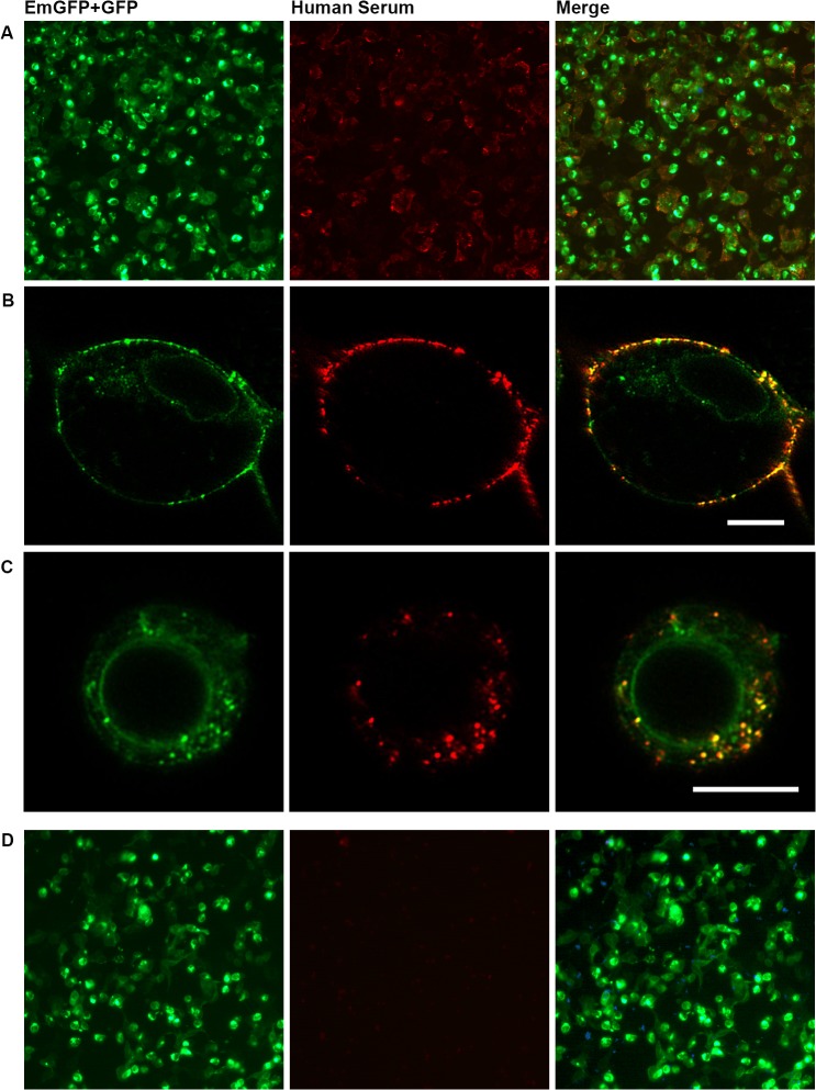
Fig 1. Immunofluorescence CBA with HEK293A cells transiently overexpressing functional NMDAR tagged with green fluorescent proteins.
Staining pattern with NMDAR antibody positive (A-C) and negative (D) serum. HEK293A cells were transiently transfected to overexpress EmGFP-tagged NR1, NR2A and GFP-tagged NR2B, incubated with diluted human serum and NMDAR antibodies were visualized by a Cy3-conjugated secondary antibody and counter-stained with DAPI to detect dead cells (left column: green fluorescence/EmGFP+GFP; middle column: red fluorescence/Cy3; right column: overlay of EmGFP/GFP, Cy3 and DAPI (A+D)). (B)+(C) Images show colocalization of NMDAR and serum NMDAR antibodies at high magnification (scale bars: 10 μm). (B) NMDAR antibodies bound to surface of cells. (C) Bound NMDAR antibodies internalized by the cells. CBA = cell-based assay. DAPI = 4’,6-diamidino-2-phenylindole. (Em)GFP = (emerald) green fluorescent protein. NMDAR = N-methyl-D-aspartate receptor.

