Figure 1.
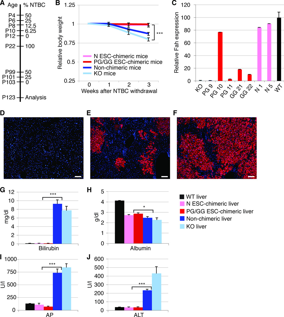
Hepatocytes derived from PG/GG ESCs rescue Fah-deficient mice from liver failure. (A) NTBC withdrawal protocol. (B) Body weight curves of PG/GG- and N ESC-chimeric, non-chimeric, and Fah-deficient (KO) control mice off NTBC. The slightly higher weight loss observed in the KO mice as compared to non-chimeric mice is most likely due to differences in mouse strain background (129S4 vs. 129S4 X C57BL/6, respectively). (C) qRT-PCR shows Fah gene expression levels 3 weeks after NTBC withdrawal (except PG 11 that was on NTBC) relative to wild-type (WT) mice. (D–F) Fah immunostaining (red) on liver sections of a non-chimeric mouse (D), and of chimeric mice with ~20% (E) or ~90% (F) liver repopulation. Nuclei are stained with DAPI (blue). (G–J) Plots showing serum concentrations of bilirubin, albumin, alkaline phosphatase (AP), and alanine aminotransferase (ALT) in adult mice 3 weeks after NTBC withdrawal. Error bars represent average relative to initial body weight ± SE for 4 N ESC-chimeric, 3 PG/GG ESC-chimeric, 4 non-chimeric, and 8 KO mice in (B), additional 3 WT mice in (G–J), and 1 WT, 2 N ESC-chimeric, 4 PG/GG ESC-chimeric, 1 non-chimeric, and 1 KO mice in (C). *P < 0.05, ***P < 0.0001. Size bars = 100 µm.

