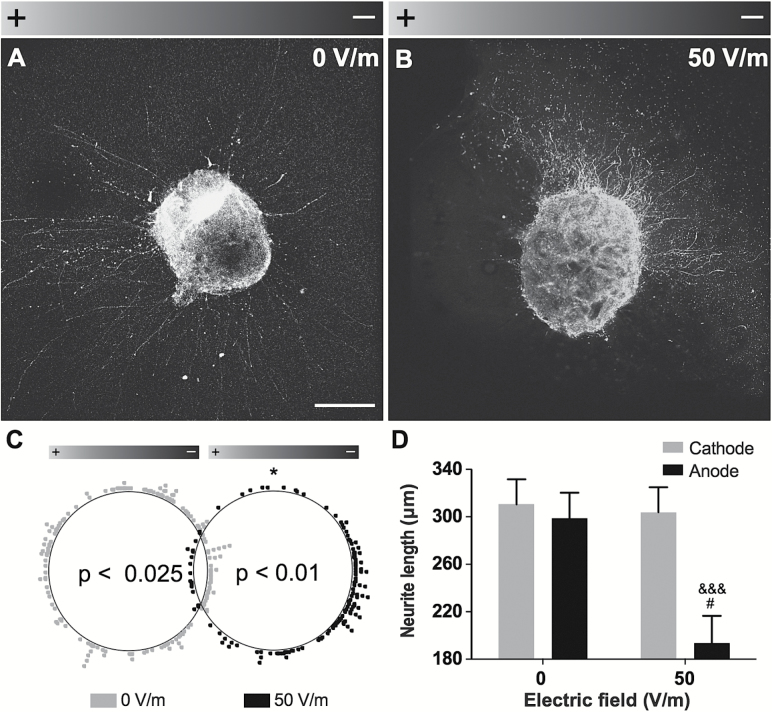
Figure 3.
DCEF effects on dopaminergic explants. (A-B) Photomicrographs of VTA explants obtained from E14.5 Pitx3-GFP transgenic mice in control condition (0V/m) or following a 24-hour exposure to a 50-V/m DCEF (n=3 explants per condition). Scale bar: 200 µm. (C) Polar scatterplot depicting the orientation of neurite outgrowth for each condition, where each dot represents the end point of a single neurite. A significant orientation shift towards the cathode was observed in the 50-V/m condition. (D) The total neurite length, as measured within the delineated quadrants corresponding to the anode and the cathode, was also significantly decreased following a 50-V/m DCEF application. Values are expressed as LS-means ± SEM. Statistical analyses were performed using the (C) Kuiper’s Test of Uniformity, (D) the Mann-Whitney following log transformation. *P<.05 vs 0V/m; # P<.05 vs anode, 0V/m; &&& P<.0001 vs Cathode, 50V/m. DCEF, direct current electrical field; LS, least square; V/m, Volt/meter.

